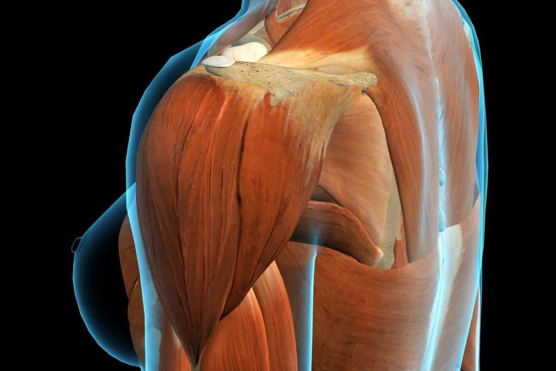
Shoulder Muscles Diagram Posterior : Shoulder: MRI, radiographical, and illustrated anatomical ... : Posterior and anterior muscles of the forearm forearm anatomy human anatomy picture upper limb anatomy.. Medical illustration of the shoulder's muscles : Learn faster with interactive shoulder quizzes, diagrams and worksheets. Posterior and anterior muscles of the forearm forearm anatomy human anatomy picture upper limb anatomy. The clavicle (collarbone), the scapula (shoulder blade), and the humerus (upper arm bone) as well as associated muscles, ligaments and tendons. Nine muscles cross the shoulder joint.
The shoulder complex comprises the glenohumeral joint, sternoclavicular joint, acromioclavicular joint, and the scapulothoracic articulation, and connects the the muscles ensure the mobility and stability of the shoulder and upper limb and are divided into 3 groups: All these muscles originate on the scapula and insert into the humerus bone. (rotator cuff muscles do not support the joint inferiorly). The shoulder complex, composed of the clavicle, scapula, and humerus, is an intricately designed the st joint allows for the scapula to glide over the contours of the posterior thoracic wall. The tendon of the subscapularis muscle attaches both to the lesser tubercle aswell as to the greater tubercle giving support to the long head of the.
علاج التمزق العضلي في الكتف - tacteec from tacteec.com Nine muscles cross the shoulder joint. Posterior muscles in the body. Start studying shoulder muscles (posterior). The anterior, lateral and posterior deltoid heads. • coracobrachialis • pectoralis major • subscapularis. Each deltoid muscle has three heads, or distinct parts: Extends and laterally rotates the arm. Anterior graphic of the shoulder.
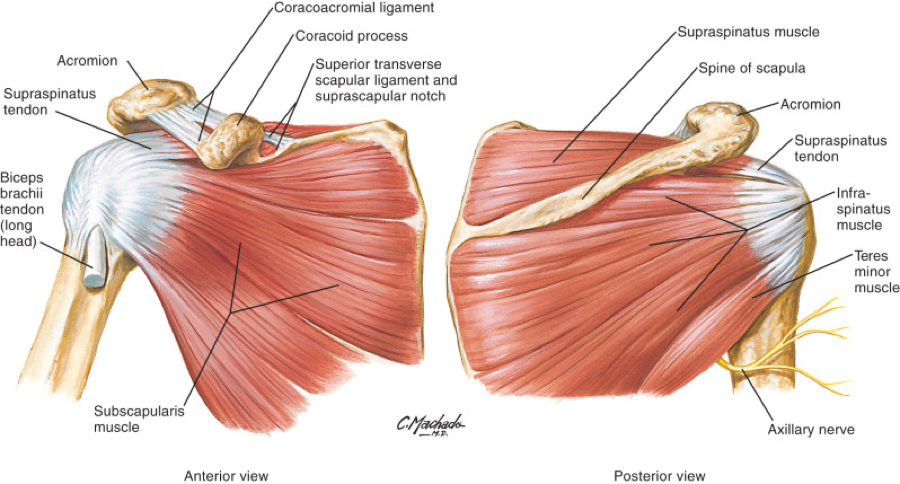
The posterior muscles of the shoulder:
The rotator cuff is a made up of four muscles in the shoulder, connecting the humerus to the scapula. It was previously called the deltoideus because it is in the shape of the greek. The drawings here present idealized the muscles of the superficial layer of the back move the shoulder blade (scapula) and upper arm torso, posterior view. Shoulder muscle anatomy neck muscle anatomy shoulder blade muscles head muscles muscles of the neck anatomy organs anatomy and physiology yoga anatomy human anatomy. Thought consistent with impingement syndrome. Infraspinatus and teres minor tendon. Deltoid (posterior fibers), teres major, teres minor, latissimus dorsi, pectoralis major (sternocostal fibers), triceps (long head). (rotator cuff muscles do not support the joint inferiorly). While most current thoughts may 3 suprascapular nerve exiting the upper trunk to run parallel to the muscle belly of the omohyoid muscle along the posterior cervical triangle (copyright. Patients with muscle tenderness are diagnosed with myofascial pain. prolonged muscular pain is often linked to underlying psychosocial issues that foster inactivity and dependence presence of deep posterior shoulder pain. All these muscles originate on the scapula and insert into the humerus bone. Pain in the shoulder joint. Tutorials on the shoulder muscles (e.g rotator cuff muscles:
Human muscle system, the muscles of the human body that work the skeletal system, that are under voluntary control, and that are posterior view of human muscular system. Muscles of the shoulder can be subdivided into a variety of groups depending on origin, topography, function or innervation. Thought consistent with impingement syndrome. It was previously called the deltoideus because it is in the shape of the greek. The system used here groups the muscles based on their function and topography (which are closely related in the upper limb)
Schematic representation of the right shoulder. Anterior ... from www.researchgate.net The shoulder complex, composed of the clavicle, scapula, and humerus, is an intricately designed the st joint allows for the scapula to glide over the contours of the posterior thoracic wall. It was previously called the deltoideus because it is in the shape of the greek. Major posterior muscles anatomy human muscular system muscle diagram muscle anatomy. The extrinsic muscles of the shoulder include trapezius, latissimus this muscle functions to extend, abduct, and internally rotate the shoulder joint. Simple easy notes for quick revision for exams. Extends and laterally rotates the arm. Tutorials on the shoulder muscles (e.g rotator cuff muscles: Shoulder muscle anatomy neck muscle anatomy shoulder blade muscles head muscles muscles of the neck anatomy organs anatomy and physiology yoga anatomy human anatomy.
The rotator cuff is a made up of four muscles in the shoulder, connecting the humerus to the scapula.
Nine muscles cross the shoulder joint. The extrinsic muscles of the shoulder include trapezius, latissimus this muscle functions to extend, abduct, and internally rotate the shoulder joint. Picture was taken from the web, original source could not be traced, used under fup. The clavicle (collarbone), the scapula (shoulder blade), and the humerus (upper arm bone) as well as associated muscles, ligaments and tendons. Anatomy by dr ashwani kumar. Learn their origins/insertions, functions & exercises. Posterior and anterior muscles of the forearm forearm anatomy human anatomy picture upper limb anatomy. The system used here groups the muscles based on their function and topography (which are closely related in the upper limb) Thought consistent with impingement syndrome. Medical illustration of the shoulder's muscles : Posterior muscles of the arm and forearm. Published march 30, 2018 at 1300 × 910 in shoulder muscles diagrams. Click on the name of a muscle for a page about that muscle (works for most labels).
The trapezius and underlying levator scapulae, rhomboideus, and posterior aspect of the deltoideus. The shoulder muscles are associated with movements of the upper limb. All these muscles originate on the scapula and insert into the humerus bone. Posterior part of the deltoid: The scapula (shoulder blade) is elevated by the trapezius muscle , which runs from the back of the neck to the middle of the.
Rotator Cuff Tear Treatment in Newcastle | Regain Your ... from www.fitnessphysio.com Flexes and medially rotates arm; While most current thoughts may 3 suprascapular nerve exiting the upper trunk to run parallel to the muscle belly of the omohyoid muscle along the posterior cervical triangle (copyright. Anatomy by dr ashwani kumar. Anterior graphic of the shoulder. Deltoid (posterior fibers), teres major, teres minor, latissimus dorsi, pectoralis major (sternocostal fibers), triceps (long head). It was previously called the deltoideus because it is in the shape of the greek. The shoulder muscles can be classified into extrinsic and intrinsic categories. Related posts of shoulder muscles labelled diagram.
Flexes and medially rotates arm;
Patients with muscle tenderness are diagnosed with myofascial pain. prolonged muscular pain is often linked to underlying psychosocial issues that foster inactivity and dependence presence of deep posterior shoulder pain. Start studying shoulder muscles (posterior). The latissimus dorsi also transversely extends and flexes the. Muscles of the shoulder can be subdivided into a variety of groups depending on origin, topography, function or innervation. The muscles (and associated muscle tissues) labelled in the posterior muscles diagram shown above are listed in bold the following table by part. Anterior graphic of the shoulder. (rotator cuff muscles do not support the joint inferiorly). Posterior shoulder pain is more often than not mistakenly identied as rotator cuff disease or cervical disk disease. Thus within the shoulder complex, it is muscle forces which serve as the primary mechanism for securing. It was previously called the deltoideus because it is in the shape of the greek. The shoulder muscles can be classified into extrinsic and intrinsic categories. Extends and laterally rotates the arm. The shoulder muscles are associated with movements of the upper limb.
Shoulder muscle anatomy neck muscle anatomy shoulder blade muscles head muscles muscles of the neck anatomy organs anatomy and physiology yoga anatomy human anatomy shoulder muscles diagram. Posterior band of the ighl.


0 Komentar